As part of the CERT project, we also became involved in the design and testing of x-ray lenses, or concentrators, in the hope of increasing the x-ray dose delivered to a tumor without increasing the dose to normal tissue. X-rays are notoriously difficult to focus, since they tend to go through things rather than be reflected or refracted. However, using Bragg reflection one can indeed reflect x-rays, and there is some hope of making a concentrating x-ray mirror using this effect. But it’s much harder to use this to make an x-ray lens than it might be to make an optical lens using a mirror. The problem is that the x-ray reflection only occurs at one specific angle of incidence for a given x-ray energy; with an optical mirror a light ray of any angle of incidence will be reflected. A flat mirror would only reflect a very small subset of the x-rays impinging on its surface.
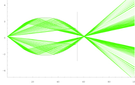
The problem of designing an x-ray concentrator comprises two parts: selecting a material, and forming that material into the correct shape. The material part is relatively straightforward: conventional silicon wafers offer a large area of single crystals that have an atomic spacing suitable for meeting the Bragg condition for the range of x-ray energies we were considering. In deciding on the form of the reflecting surface we relied on a remarkable property of the mathematical shape called a logarithmic spiral. It has the characteristic that any ray from the origin of the coordinate system to the surface of the log spiral will meet that surface at a constant angle. So an x-ray source placed at the origin will result in all of its rays hitting the surface at the desired Bragg angle. Unfortunately the reflected rays (which also leave the surface at the same angle) do not come together at an exact focus, but it’s close enough that an effective concentrator can be constructed.
In addition to performing the design of optimal shapes (using Mathematica), which also accounted for the non-monochromatic spectrum of the source, we also embarked on a program of testing our results. We had a mechanical device made that allowed a long strip of silicon to be precisely bent (under computer control) into a variety of curved shapes. Then we constructed a multi-axis computer-controlled motion control setup to vary the position and orientation of an x-ray source, the silicon reflector, and an imaging x-ray detector. Everything was automated using motor drives and encoders, mostly acquired through eBay since the budget was very tight. PC software (a mixture of C and Tcl/Tk) controlled the experiments and collected data. With so many degrees of freedom it was imperative that everything run for hours without operator intervention (or even presence). Which it did.
The image above shows a concentrator made out of segments of different log spiral curves. The focus is much better than a single log spiral.